
What is an OCT?
Optical coherence tomography (OCT) is a non-invasive medical imaging technique. It has revolutionised the diagnosing and managing of ocular conditions. Once when only hospitals would have OCT’s, it is now readily being used in high street practices. As a Locum, it is important to understand, interpret and diagnose using an OCT, because some stores make it compulsory to use an OCT during a routine sight test.
Hears a little guide of all the different scans, we won’t bore you with the science but what to look out for.
Step 1
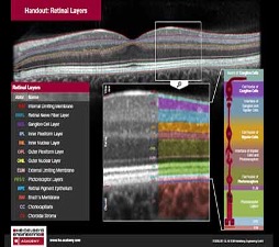
Understand all the retinal layers of the eye. OCT is one of the tools to help aid your diagnoses, and its primary role is to show you where things are changing. Use this and your ocular knowledge and another test to understand what’s happening. So back to basic let’s look at the layers.
Step 2
OCT is based on low-coherence interferometry. OCT images are generated by measuring the echo time lag and the intensity of back-scattered light. Light in an OCT system is broken into two arms: A sample beam (containing the item of interest), a reference beam (usually a mirror).
This reflectivity profile is called an A-scan and contains information about the spatial dimensions and location of structures within the item of interest. A cross-sectional tomography (B-scan) may be attained by laterally combining a series of these axial depth scans (A-scan). OCT combines lots of A-scans to build B-scan images; B-scans are subsequently used to produce 3D maps or cubes.
A-scans identify boundary locations as the intensity peaks in each scan and link the feature points to form a smooth and continuous boundary.
B-scan produces high-resolution images and can be beneficial in assessing the retina in more detail.
C-scan is a B-scan over some time. Eg 3 months’ intervals.
Different types of scan
Raster scan
A scale-like is a horizontal cross-section of the retina (A-scan). Raster scan is multiple-scale lines, separated by a set distance, larger the gap. The less detailed the scan is, with a higher chance of miss a lesion but is quicker, closer the distance the more detail. Most OCT Raster scan has closer A-scans nearer to the fovea and separate as the scan moves to the periphery.
Volume scan
A volume scan is built up of multiple B-scans. It is a series of horizontal and vertical raster scans combined to create a cube of images. Volume scans allow for a more extensive area of the retina to be scanned without the patient having to change their fixation. This is ideal when screening for abnormalities, or to build up an overall impression of an area of interest.
A radial line
A radial line scan directs the OCT (A-scan) beam radially, like a circle plot. A series of 6-12 radial line scan through the macula.
Advantage- high resolution and covers a large area of the macula.
The disadvantage, potential miss lesion due to large lines in the para-foveal area.
Once you have conducted the OCT scan we need to examine the scan. Also, remember an OCT is one of the tools to help with your diagnoses. In a combination of symptoms, Volk examination, visions and another test you can make a better management plan. OCT’s have a retinal thickening map, with a green, amber and red colour chart to help with glaucoma management. The average retinal thickness is 22216m and 260 12m for the central macula area.
Use this guide to know what scan to do, dependent on what you’re looking for. Each scan help you find your answer.
For more information on Locumotive make sure to visit our website. Also, make sure to read our other blogs too.
![]()

Leave A Comment